The health industry is evolving almost daily with new and enhanced practices, advanced patient experiences and newer treatment outcomes. The dental industry is also evolving similarly, where state-of-the-art technologies, precise software, 3D printing, and more have been digitised at their best.
In this blog, let’s look at digital occlusion analysis and how fruitful it is for precision dentistry.
What is Digital Occlusion Analysis?
Occlusion analysis, in simple language, is the occlusal relationships between your upper and lower teeth. It refers to the systematic evaluation of a patient’s bite, that is, the relationship between their upper and lower teeth when they close their jaws. It’s crucial for malocclusion analysis or diagnosing dental disorders like improper alignment of teeth), bruxism (teeth grinding), and Temporomandibular Joint Dysfunction (TMJD).
A thorough occlusion analysis can facilitate the detection of occlusion-related issues at an early stage, making it a vital part of a comprehensive dental examination.
Traditionally, the occlusion analysis involved wax bites, articulating paper, and pressure indicator paste. These methods served the purpose but often needed more precision and clarity as the final citation required the dentist’s subjective interpretation, which was not similar from one professional to the other. Furthermore, they provided limited information, making it challenging to pinpoint the location and degree of occlusion discrepancies accurately.
Digital occlusion analysis uses computerized technology and high-resolution sensors to evaluate bite forces, timings, and balance, offering unparalleled precision and consistency.
Why is Digital Occlusion an Important Tool of Digital Dentistry?
Digital technology has drastically shifted the dental landscape, offering precision, efficiency, and patient comfort. In occlusion analysis, these advancements have opened up a whole new world of techniques that enhance our diagnostic capabilities and treatment planning.
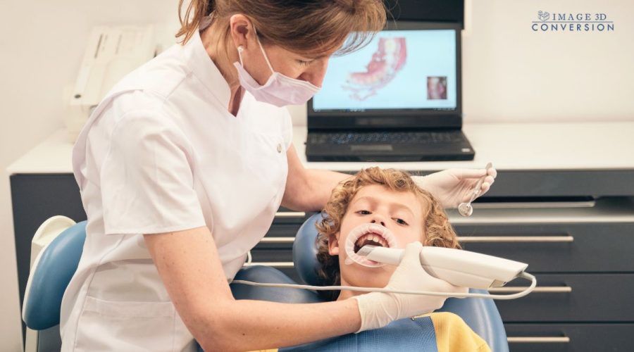
Intraoral Scanners are one of the most significant advancements in digital dentistry. Replacing traditional impression techniques, intraoral scanners provide a seamless, fast, and highly accurate alternative. The scanner projects a light source onto the oral structures and records the reflections to create a continuous stream of 3D digital images, generating a comprehensive digital impression.
Benefits It Offers
Accuracy and precision are the cornerstones of any medical analysis, and dental occlusion is no exception. Digital occlusion analysis surpasses its traditional counterparts by offering unprecedented detail and precision. Dentists can devise more accurate and personalized treatment plans with the detailed, dynamic information that digital occlusion analysis provides. They can also predict potential complications and address them proactively. It also offers:
- Enhanced Precision: By digitally mapping the contact points between teeth, dentists can now identify every imbalance that traditional methods may have missed.
- High-resolution Imaging: Digital occlusion analysis tools, such as intraoral scanners and computerized occlusal analysis systems, provide high-resolution images, capturing minute details of a patient’s bite. This leads to more accurate diagnoses and efficient treatment planning.
- Effective Case Presentation: Visuals play a crucial role in case presentations. The digital 3D models and simulations help patients better understand their dental conditions and proposed treatments, thus facilitating informed consent and improving patient satisfaction.
- Objective Data: Digital occlusion analysis gives quantifiable data, allowing for accurate interpretation, minimizing subjectivity and enhancing the accuracy of the diagnosis.
- Improved Patient Comfort: The digital process is non-invasive and more comfortable for patients, enhancing their overall experience.
- Eco-friendly: Digital impressions are a greener alternative, reducing the need for disposable impression materials and trays.
- Efficient Communication with the Dental Laboratory: Digital occlusion analysis effectively addresses the communication gap between the dentist and the dental laboratory, especially regarding occlusion issues.
- Digital Data Transfer: The 3D digital impressions and occlusal data can be shared instantly with the dental laboratory, reducing the turnaround time.
- Consistency in Results: The accurate digital data ensures consistency between the dentist’s treatment plan and the laboratory’s execution, resulting in more successful treatment outcomes.
Digital Occlusion Analysis: Future Scopes and More
Compact Devices: Compact, handheld intraoral scanners are becoming increasingly popular, offering convenience and flexibility in occlusal data capture.
Real-time Analysis: Some of the latest occlusal analysis systems provide real-time data, allowing instant interpretation and application during patient consultations.
Integrated Systems: More dental practices are adopting fully integrated systems, allowing seamless data sharing and collaboration between different aspects of patient care.
Automated Analysis: Artificial Intelligence (AI) and Machine Learning (ML) are opening up new avenues in digital occlusion analysis. AI and ML can help automate the interpretation of occlusal data, identifying patterns and anomalies more quickly and accurately than manual analysis.
Predictive Modelling: ML algorithms can be trained to predict future occlusal changes, aiding preventive dentistry and early intervention.
Enhanced Training: AI-powered simulation models can be effective training tools, helping dental professionals improve their diagnostic and treatment planning skills.
Conclusion
As we look ahead, it’s clear that the future of bite analysis and treatment planning is increasingly digital, data-driven, and precise. With AI and Machine Learning adding another layer of sophistication, the potential for predicting occlusal changes and personalized preventive dentistry is now within our grasp.
As we continue this digital transformation journey, we are shaping a future where the union of technology and dental expertise leads to superior patient care.
FAQs
Digital occlusion analysis uses computerized technology and high-resolution sensors to evaluate bite forces, timings, and balance, which is much more precise than traditional methods.
It’s precise, accurate and non-invasive.
An occlusion analysis can be done using digital techniques for between half an hour.
Yes, digital occlusion analysis is highly beneficial for prosthodontics in many ways. It enhances precision in diagnosing and treating occlusal issues by providing detailed, objective data on bite forces and timing, which traditional methods like articulating paper cannot offer.
Digital occlusion analysis typically involves the use of various technologies. Some of the key technologies include:
– Articulating Paper
– Computerized Occlusal Analysis Systems
– Digital Scanners
– CAD/CAM Technology
– Cone Beam Computed Tomography (CBCT)
– Electromyography (EMG)
– Pressure Mapping Systems
– Virtual Articulators
References
Mihaylova, A., Yordanova, M., & Yordanova, S. (2023). “T-Scan Novus System Application—Digital Occlusion Analysis of 3D Printed Orthodontic Retainers.” Applied Sciences, 13(14), 8111.
Gu, D.A., Miao, L.Y., & Liu, C. (2022). “Application of Digital Occlusion Analysis System in Stomatological Clinical Medicine.” Austin Journal of Dentistry, 9(1), Article ID 1164.
Alex, G., & Polimeni, A. (2006). “Comprehensive Dentistry: The Key to Predictable Smile Design.” AACD Monograph, 15-20.
van der Bilt, A., Tekamp, A., van der Glas, H., & Abbink, J. (2008). “Bite Force and Electromyography during Maximum Unilateral and Bilateral Clenching.” European Journal of Oral Sciences, 116(3), 217-222.
Lundeen, H.C., & Gibbs, C.H. (1982). “Jaw Movements and Forces during Chewing and Swallowing and Their Clinical Significance.” In Advances in Occlusion (pp. 23-45). Boston: John Wright.


